Dental Implants
Evaluating Your Professional Options for Care
(Continued)
Insufficient Bone — “Regeneration” In Our Generation
Sufficient bone volume for implant placement is vitally important to proper tooth placement resulting in both the most natural-looking and properly functioning tooth. Today there is general scientific agreement supporting the concept that when a tooth is removed a bone graft placed into the extraction site will minimize inevitable melting away of bone or “resorption.” Maintaining “bone volume” following removal of a tooth will facilitate implant placement in the best possible position.
Understanding the principles of wound healing now allows for regeneration of bone to occur using a variety of techniques. Most include opening the gingival (gum) tissues to expose the bone and then augmenting the existing or remaining bone by adding bone grafting materials to it. Healing of the bone can be enhanced by the utilization of membranes which cover the grafts and act like little subterranean band-aids to “guide bone regeneration.” Along with other biologically active molecules (found normally in the body) these techniques promote and enhance healing. In addition, excellent techniques exist for replacing and adding gingival gum tissues.
These surgical procedures are generally carried out by a periodontist or oral surgeon skilled and experienced in these techniques, especially in advanced situations. When creating new bone for implant placement, particularly in the upper jaw where sinuses are involved and bone grafting is necessary, these procedures are more predictably carried out in the hands of a specialist or a general dentist who has taken special and advanced training.
 |
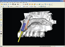
| Figure 3: Using CAT scan technology, a dentist can verify that there is sufficient bone to place an implant in the right location for an aesthetically pleasing crown. |
| Photo courtesy of Materialise Dental Inc. |
Implant Placement and Positioning
Sometimes described as “top down treatment planning,” the teeth to be replaced are recreated in a wax model form by a dental laboratory technician. The idea is then to establish the position of the underlying bone and to make sure the implant(s) is properly aligned (down) with the wax tooth form (top). The implant position can then be predetermined using a combination of specialized radiographs (x-rays) and imaging technology to assure success and in the process avoid major structures like nerves and air sinuses [Figure 3].
From this information surgical guides are made to assist the surgeon in precise implant placement; this in turn will assure the restorative dentist (general dentist or prosthodontist) that a crown will fit in the right position. If the bite will not accommodate implant placement, orthodontic treatment (braces carried out by an orthodontist) may be necessary to reposition teeth.
This process is analogous to the scuba diving adage, “Plan the dive and dive the plan.” A lot of preparatory work goes into initially deciding where an implant is going to be placed long before the actual surgery.
Finally, even with all the appropriate diagnosis and preparatory work, it's not a slam dunk — surgical know how does count. Surgical technique is in part an art, dependent upon proper knowledge, training and experience that can take years to acquire. It really comes down to this: every expert is an artist in his/her own field. Working on his/her particular canvas with all the appropriate information and experience at hand, the surgeon creates a work of art using materials with which he/she is most familiar.

