
CAT Scans In Dentistry
A new standard for the 21st century
 Dear Doctor,
My orthodontist wants me to get a CAT scan for my son before he gets braces. Why a CAT scan and is it worth the money?
Dear Julian,
If a picture is worth a thousand words, what are a thousand, or even a million pictures worth? Imagine how much more you could learn about objects if you could actually see them from the inside, life size and in color. Viewing a well-painted landscape is nothing like living and walking through a landscape. Studying a photograph of a house does not compare to walking through the house room-by-room. And so it has been in the art and science of healthcare since Roentgen first discovered the ability to image the body with radiographs (x-ray pictures). But all x-rays — just like paintings and photographs — have been two dimensional (2-D), until now.
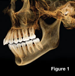
CBCT scanning or Cone Beam Computed Tomography (“compute” – to determine or calculate by mathematical means; “tomo” – slice; “graph” – to write) now provides images in three dimensions or “3-D” as it is more commonly referred. CBCT is the latest type of Computer Assisted Tomograph or CAT scan. For medical and dental professionals, it really does provide the ability to see and experience the body from the inside; thus changing the way diagnoses can be made. And the implications for treatments are staggering [Figure 1].
It really combines the best of both worlds. A Cone Beam (CB) uses digital geometry to generate a three-dimensional image of the inside of an object from a large series of two-dimensional images. The CB actually refers to a spiral beam of x-rays that emanate from and create a cone shaped beam, which is then caught on detectors.
Envision the way layers make up your favorite multi-layered cake and that's how the CBCT scanner builds up the images, by layering them one-by-one on top of each other. Now think of the slices you cut out of the cake to eat it — the thinner the slices the more you see of the cake's ingredients, such as finely diced nuts or berries. Using this analogy, these many images make up the results of a 3-D scan.
 |
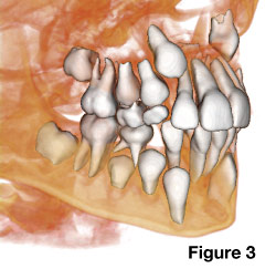 |
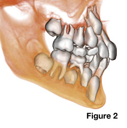
| Figure 2 and 3: An orthodontist will use these CAT scan images to plan treatment based on growth and development of your child. Relating the size of the permanent teeth, when they will come into the oral cavity, what position they will likely erupt into and if they need to be extracted, are just some of the questions that can accurately be determined with the use of 3-D radiography (x-ray images). |
| Figures 1, 2 and 3 provided by Anatomage, Inc. |
Coming To An Office Near You
Believe it or not, CBCT greatly benefits patients and makes dentistry more fun to practice. For these reasons, among others, the use of CBCT in dentistry is rapidly increasing. The following list highlights some of these areas by specialty:
- Dentists and dental specialists using CBCT technology today have the ability to visualize true three-dimensional anatomy of the teeth, jaws and facial skeleton, as well as other vital structures of the head and neck region in precise and extreme detail. CBCT enhances diagnosis and treatment planning; thus patients benefit greatly.
- Orthodontists and Pediatric dentists can examine the growth stages of skeletal structures and developing teeth using CBCT technology as well as using it for monitoring health, identifying disease and planning interceptive strategies to direct growth in the best possible direction [Figure 2 and 3].
- Cosmetic dentists and prosthodontists (tooth replacement specialists) use CBCT technology for precisely imaging the teeth, the bite and Temporo-Mandibular Joints (TMJ). They also use it for analyzing and rebuilding teeth and bites with crowns, implants and more.
- Oral surgeons use CBCT to find disease, determine fractures due to trauma, look for impacted or missing teeth and more. The position of impacted wisdom teeth and their proximity to nerves and sinuses can be precisely determined from a 3D CBCT scan.
- Endodontists (root canal specialists) find CBCT scans are detailed enough to look into root canals less than a millimeter in width, to diagnose and treat abscessed teeth, and even to identify tiny accessory canals that do not show easily on conventional 2D x-rays.
- Periodontists (gum specialists) can use CBCT technology to assess bone loss gum attachment levels in greater detail to make more precise diagnoses. And as a result, they are better equipped to perform regenerative surgical treatments to prolong the life of teeth.
- Some dental specialists treating sleep disorders such as snoring and Obstructive Sleep Apnea (OSA; a condition in which airway or windpipe blockage and choking occurs while sleeping) use CBCT to obtain clear 3D images of the airway. Analysis of these living images during sleep makes treatment strategies more realistic.
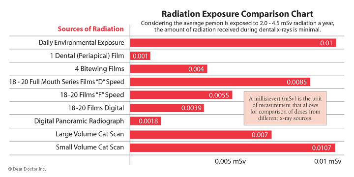 |
| Figure 4: The radiation doses from CBCT are low when compared to conventional CT, but are significantly higher than conventional dental radiographic x-ray techniques. It is useful to compare these doses for conventional films used in dentistry. Click to enlarge |
How Do They Compare
CBCT technology delivers a higher dose of x-rays than a typical 2D panoramic radiograph. It delivers a lower dose than a typical digital standard 18 film full mouth x-ray series. Effective doses from large volume CBCT scanners have been reported ranging from approximately 70 µSv to 1070 µSv [Figure 4]. Surprisingly scans using a smaller field of view actually have a higher dose but provide much better detail of the dental anatomy. Generally the doses are significantly lower than conventional medical CT scanners.
The Cone Beam Advantage
One low-dose CBCT scan generates an extremely accurate assessment of bone quality, quantity, as well as a better understanding of associated anatomical relationships in three dimensions. CBCT also allows precise treatment planning to occur before, rather than during treatment delivery. Imagine being able to map in a virtual computer environment an individual's underlying anatomy and dental problems with precise accuracy prior to starting his or her treatment. The confidence generated from CBCT technology improves efficiency, work quality and most importantly treatment predictability.