
Sinus Surgery
Creating Bone For Dental Implants Out Of Thin Air
(Continued)
Imaging The Sinus
First, the shape, location, and health of the sinus need to be assessed before surgery. X-rays will be required as part of the evaluation to determine the extent and type of surgery necessary. A panoramic radiograph (x-ray) is used to provide a two-dimensional image of the jawbones and skull including the sinuses. This may further indicate the need for a detailed three-dimensional image, a CT scan (C – Computed; T – Tomogram from “tomo” – slice), to provide accurate information regarding the shape and volume of the sinus for surgical assessment. The importance of these diagnostic images is based upon the surgeon's clinical judgment and experience.
|
Sinus Surgery A Look From Start to Finish |
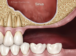 |
| Side view of the sinus showing insufficient bone to replace the missing upper posterior teeth. |
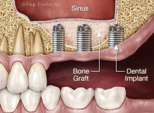 |
| Lifting of the sinus membrane creates space for bone grafting which increases the volume of bone for placement of dental implants. |
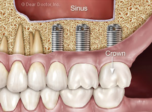 |
| After 6 to 7 months of healing, the dental implants are restored with implant crowns which look and function like natural teeth. |
How It's Done: Surgical Techniques
The surgical procedures are performed from inside the mouth in the area just above the missing back teeth. They are generally carried out under local anesthesia by numbing the area as is done for a routine filling. Some patients require addition sedation or anti-anxiety medication, which can either be administered orally, by mouth, or by intra-venous injection (“intra” – into: “venous” – vein). These options should be discussed with your dentist.
Sinus Osteotome Technique
If there is sufficient bone to stabilize an implant but not fully anchor it, a more minor sinus augmentation can be done. This is achieved by accessing the sinus membrane directly through the site for implant placement and simultaneously placing the implant(s). It is performed using instruments called osteotomes (“osteo” – bone; “tome” – shaping), which allow the sinus membrane to be very gently accessed and raised through the implant site. These surgical tools also give this procedure its name, the Summer's Sinus Osteotome Technique.
The Lateral Window Technique
If there is very little or no bone available, then a small window (about the length of two or three teeth) is created from within the mouth accessing the cheek-side bony wall of the sinus. This is known as a lateral (side) window technique. A small incision is first made inside the mouth to expose the sinus wall. Next, a window is made in the bone to expose the sinus membrane, which is carefully and gently elevated in the area of the teeth to be replaced. The grafting materials are placed under the sinus membrane and the window is neatly closed. The grafted material is left to be covered by and/or converted into the body's own bone, a process taking six months or more before implants can be placed. If there is sufficient bone to stabilize the implants, they can be placed simultaneously with the graft, thus saving time and avoiding a second surgical procedure.
 |
| Manufactured bone grafting materials simplifies surgical procedures. |
Grafting Materials: Since 2003 bone replacement grafts, known as xenografts (“xeno” – strange or foreign, from a different animal species; “graft” – a surgical procedure to transplant tissue) have been shown to work as well, if not better than human bone autografts (“auto” – self, from the same person). In fact our best scientific evidence shows implant success rates of 98% in sinuses grafted with xenograft material. The most well researched bone substitute grafting material in use today is bovine (cow) bone. It has a higher success rate than implants placed in the natural bone of the area.
It is important to note that all grafting materials used today are approved by the Food and Drug Administration (FDA) and must be prepared according to their guidelines. They are specially treated to render them completely sterile, non-contagious, and free of rejection factors. For the most part, bone grafts act as scaffolds that the body replaces with its own bone.
Enhancers: Current research is focusing on enhancers or growth factors, substances or molecules mostly protein in nature, that may both speed up and enhance the quality of bone being grown or generated. These include Bone Morphogenetic Protein (BMP; “morpho” – shape; “genesis” – to grow) and Platelet-Derived Growth Factors (PDGF). (Note: Platelets are tiny, little, “plate-like” structures floating in the blood, that allow clotting and in so doing release growth factors that initiate and promote healing.) While both of these substances occur naturally in the body, they can be synthesized today in the laboratory. These factors hopefully will produce more bone and/or do it in less time. However, there are currently few studies available that validate their universal use.
Closing The Window: When the window to access the sinus membrane is closed, most evidence-based research points to the fact that healing is enhanced. This is accomplished most often by placing a membrane over the window which is then covered by the gum tissue and oral mucosa. This thin membrane functions like a band-aid; however, its magic is that it dissolves and does not require another surgery to remove.